The left subclavian and left common carotid arteries arise independently from the aortic arch but otherwise follow a similar pattern and distribution to the corresponding arteries on the right side (see Figure 20.23). Each subclavian artery supplies blood to the arms, chest, shoulders, back, and central nervous system. It then gives rise to
Heart (right and left atrium): Anatomy and function | Kenhub
Label the major blood vessels of the pulmonary and systemic circulations; … veins share the same general features, but the walls of arteries are much thicker because of the higher pressure of the blood that flows through them. … Figure 20.28 Arteries of the Thoracic and Abdominal Regions The thoracic aorta gives rise to the arteries of the
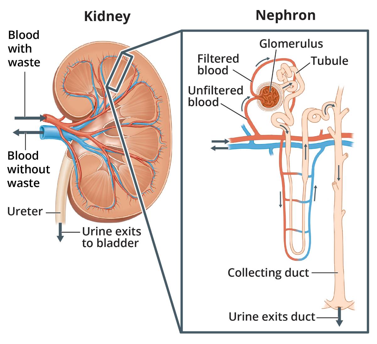
Source Image: niddk.nih.gov
Download Image
Figure 2. (a) Arteries and (b) veins share the same general features, but the walls of arteries are much thicker because of the higher pressure of the blood that flows through them. (c) A micrograph shows the relative differences in thickness. … This type of artery is known as an elastic artery (see Figure 3). Vessels larger than 10 mm in
Source Image: nhcs.com.sg
Download Image
Body System: Cardiovascular Figure 20.8 Major Arteries and Veins of Circulation. … Sketch and label what you observe using low magnification and high magnification in the space below. Your sketch should allow you to label the following structures: artery, vein, capillary, endothelium, tunica interna, tunica media (on vein and artery only), tunica externa (on vein and

Source Image: quizlet.com
Download Image
Label The General Arteries In The Figure
Figure 20.8 Major Arteries and Veins of Circulation. … Sketch and label what you observe using low magnification and high magnification in the space below. Your sketch should allow you to label the following structures: artery, vein, capillary, endothelium, tunica interna, tunica media (on vein and artery only), tunica externa (on vein and Function and anatomy of the heart made easy using labeled diagrams of cardiac structures and blood flow through the atria, ventricles, valves, aorta, pulmonary arteries veins, superior inferior vena cava, and chambers. Includes an exercise, review worksheet, quiz, and model drawing of an anterior view (frontal section) of the heart in order to
Palpable Arteries for Pulse (Labelling Exercise) Diagram | Quizlet
Figure 20.3 Structure of Blood Vessels (a) Arteries and (b) veins share the same general features, but the walls of arteries are much thicker because of the higher pressure of the blood that flows through them. (c) A micrograph shows the relative differences in thickness. LM × 160. Circulatory system | healthdirect
_d9750aa7-7903-4ed0-936e-70d01b22e08f-5ff882.gif)
Source Image: healthdirect.gov.au
Download Image
Relationship Between Central Artery Stiffness, Brain Arterial Dilation, and White Matter Hyperintensities in Older Adults: The ARIC Study—Brief Report | Arteriosclerosis, Thrombosis, and Vascular Biology Figure 20.3 Structure of Blood Vessels (a) Arteries and (b) veins share the same general features, but the walls of arteries are much thicker because of the higher pressure of the blood that flows through them. (c) A micrograph shows the relative differences in thickness. LM × 160.

Source Image: ahajournals.org
Download Image
Heart (right and left atrium): Anatomy and function | Kenhub The left subclavian and left common carotid arteries arise independently from the aortic arch but otherwise follow a similar pattern and distribution to the corresponding arteries on the right side (see Figure 20.23). Each subclavian artery supplies blood to the arms, chest, shoulders, back, and central nervous system. It then gives rise to
/images/vimeo_thumbnails/258778395/szSQn4KVGevixf6kNh3Sw_overlay.jpg)
Source Image: kenhub.com
Download Image
Body System: Cardiovascular Figure 2. (a) Arteries and (b) veins share the same general features, but the walls of arteries are much thicker because of the higher pressure of the blood that flows through them. (c) A micrograph shows the relative differences in thickness. … This type of artery is known as an elastic artery (see Figure 3). Vessels larger than 10 mm in

Source Image: nationwidechildrens.org
Download Image
Major Veins of the Body | Overview, Anatomy & Functions – Video & Lesson Transcript | Study.com May 15, 2022The graphic shows the major arteries (in bright red) and veins (dark red) of the system. Blood from the aorta passes into a branching system of arteries that lead to all parts of the body. It then flows into a system of capillaries where its exchange functions take place. Figure 15.3.1.2 Human circulation system

Source Image: study.com
Download Image
Abdomen and digestive system diagrams: normal anatomy | e-Anatomy Figure 20.8 Major Arteries and Veins of Circulation. … Sketch and label what you observe using low magnification and high magnification in the space below. Your sketch should allow you to label the following structures: artery, vein, capillary, endothelium, tunica interna, tunica media (on vein and artery only), tunica externa (on vein and

Source Image: imaios.com
Download Image
Heart Blood Flow | Simple Anatomy Diagram, Cardiac Circulation Pathway Steps — EZmed Function and anatomy of the heart made easy using labeled diagrams of cardiac structures and blood flow through the atria, ventricles, valves, aorta, pulmonary arteries veins, superior inferior vena cava, and chambers. Includes an exercise, review worksheet, quiz, and model drawing of an anterior view (frontal section) of the heart in order to
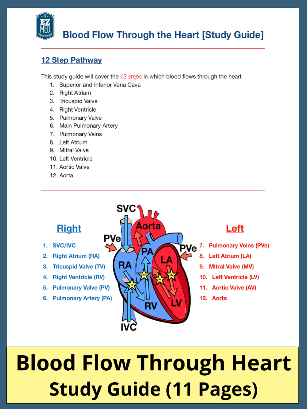
Source Image: ezmedlearning.com
Download Image
Relationship Between Central Artery Stiffness, Brain Arterial Dilation, and White Matter Hyperintensities in Older Adults: The ARIC Study—Brief Report | Arteriosclerosis, Thrombosis, and Vascular Biology
Heart Blood Flow | Simple Anatomy Diagram, Cardiac Circulation Pathway Steps — EZmed Label the major blood vessels of the pulmonary and systemic circulations; … veins share the same general features, but the walls of arteries are much thicker because of the higher pressure of the blood that flows through them. … Figure 20.28 Arteries of the Thoracic and Abdominal Regions The thoracic aorta gives rise to the arteries of the
Body System: Cardiovascular Abdomen and digestive system diagrams: normal anatomy | e-Anatomy May 15, 2022The graphic shows the major arteries (in bright red) and veins (dark red) of the system. Blood from the aorta passes into a branching system of arteries that lead to all parts of the body. It then flows into a system of capillaries where its exchange functions take place. Figure 15.3.1.2 Human circulation system
